Quick Answer: What Is the Difference Between PSG and a Home Sleep Test?
Polysomnography, or PSG, is an attended sleep study that records brain activity, sleep stages, breathing, respiratory effort, oxygen saturation, heart rhythm, body position, and muscle activity. A home sleep apnea test records fewer signals and usually cannot confirm when the patient is asleep, identify sleep stages, or detect cortical arousals.1
Home sleep apnea testing can improve access to diagnosis and may be appropriate for selected adults with suspected uncomplicated obstructive sleep apnea. However, PSG provides more physiologic context. For dentists, those differences can matter when reviewing a patient for oral appliance therapy.1
Why Sleep-Study Quality Matters to Dentists
Dentists do not diagnose obstructive sleep apnea. However, qualified dentists frequently review physician diagnoses and sleep-study reports when evaluating patients for oral appliance therapy.
The type and quality of the study can influence:
- Case selection
- Patient expectations
- Appliance selection
- Titration decisions
- Treatment follow-up
- Communication with the prescribing physician
Professional guidance supports an interdisciplinary model in which dentists screen appropriate patients, provide oral appliance therapy following a medical diagnosis, monitor dental effects, and work with physicians to confirm treatment effectiveness.2,3
Evolution of sleep diagnostics
Yet many dentists focus primarily on one number: the apnea-hypopnea index, or AHI.
Two patients may have the same AHI but very different physiologic patterns. One may experience short respiratory events with limited oxygen change. Another may have longer events, deeper desaturations, REM-related obstruction, or repeated arousals.
The number may be the same. The overnight pattern may not be.
To explore that distinction, we spoke with Ryan B. Pierce, RPSGT, a registered sleep technologist with more than 25 years of experience conducting attended sleep studies.
Ryan does not encounter sleep only through a completed report. He watches the signals develop throughout the night.
“There’s what the report says, and then there’s what actually happens in the room.”
That observation frames the central lesson for dentists: a sleep-study report reflects not only the patient’s physiology, but also the signals recorded, the conditions captured, and the way the study was conducted.
What Does PSG Measure?
An attended PSG records several physiologic systems at the same time. Depending on the study protocol, the recording may include:
- Electroencephalography: Confirms sleep and identifies sleep stages
- Electro-oculography: Helps identify rapid eye movement sleep
- Chin electromyography: Records changes in muscle tone
- Airflow sensors: Detect reductions or pauses in breathing
- Respiratory-effort belts: Measure chest and abdominal effort
- Pulse oximetry: Measures blood oxygen saturation
- Electrocardiography: Records heart rhythm
- Body-position monitoring: Identifies supine and nonsupine sleep
- Leg electromyography: Detects limb movement
The primary difference is not simply the number of sensors. PSG is attended and managed while the patient sleeps.1
A trained sleep technologist can:
- Correct displaced or failing sensors
- Identify and reduce signal artifact
- Document patient movements and behavior
- Confirm whether the patient is awake or asleep
- Observe patterns as they develop
- Help capture clinically relevant sleep positions when appropriate
Ryan explains the distinction this way:
“You’re watching it happen. You’re not guessing what happened after the fact.”
That real-time oversight helps protect signal quality. It also provides context that may be unavailable when testing is performed without a technologist present.
Why Sleep Stage and Body Position Matter
Obstructive sleep apnea does not always remain consistent throughout the night.
Respiratory events may change with:
- REM versus non-REM sleep
- Supine versus lateral position
- Sleep depth
- Alcohol or medication use
- Nasal congestion
- Night-to-night variability
Adults cycle through non-REM and REM sleep repeatedly, with longer REM periods typically occurring later in the night. Respiratory obstruction may become more pronounced during REM in some patients. Body position can also affect upper-airway collapsibility, particularly when a patient sleeps on the back.3
A study that captures little REM sleep may not reveal a patient’s REM-related pattern. Likewise, a study with little or no supine sleep may not demonstrate clinically relevant positional obstruction.
Ryan sometimes watches those differences emerge during PSG:
“If someone never goes onto their back, I may ask them to roll over for a while. That’s sometimes when you see the breathing pattern change.”
That does not mean a technologist should manufacture a diagnosis or force a patient to remain in an unnatural position. It means that testing conditions influence what appears in the final report.
Clinical takeaway for dentists
When reviewing a sleep study, look beyond the overall AHI. Consider:
- Total sleep time
- Sleep efficiency
- Amount of REM sleep
- Time spent supine
- REM-specific AHI
- Supine AHI
- Oxygen-desaturation patterns
- Central versus obstructive events
- Technical limitations documented in the report
What Does a Home Sleep Apnea Test Measure?
Home sleep apnea testing, commonly abbreviated HSAT and sometimes called HST, is an unattended study performed outside a sleep laboratory.
The signals recorded vary by system. Most devices measure some combination of:
- Airflow
- Respiratory effort
- Oxygen saturation
- Heart rate
- Snoring
- Body position
Most conventional home tests do not record the brain activity required to identify sleep stages or confirm total sleep time. They also cannot detect cortical arousals in the same way as PSG. No technologist is present to correct sensors, verify signal quality, or document events as they occur.1
Ryan describes the technology this way:
“These systems are making assumptions. They may be informed assumptions, but they are still assumptions.”
That observation should not be interpreted as a rejection of home testing. AASM guidance supports HSAT as a diagnostic option for appropriately selected adults who have signs and symptoms suggesting an increased risk of moderate to severe OSA and who do not have complicating conditions that favor PSG.1
The limitation is context. A home test may provide enough information for one patient while leaving important questions unanswered for another.
AHI Versus REI: Why the Difference Matters
PSG calculates respiratory events using confirmed sleep time. The result is reported as the apnea-hypopnea index, or AHI.
Because most home tests cannot determine actual sleep time, they usually calculate events using recording or monitoring time. This result is commonly reported as the respiratory event index, or REI.1
Consider a patient who wears a home-testing system for eight hours but sleeps for only six. Dividing respiratory events by eight hours instead of six may produce a lower event rate.
As a result, HSAT may underestimate respiratory-event frequency in some patients, particularly those who:
- Take a long time to fall asleep
- Spend substantial time awake during the night
- Have fragmented sleep
- Have mild or borderline findings
Ryan summarizes the issue directly:
“If you don’t know whether the patient is asleep, you’re starting with an estimate.”
The practical lesson is not that every REI is unreliable. Rather, AHI and REI are calculated differently and should not always be treated as interchangeable.
PSG vs Home Sleep Test: Key Differences
|
Clinical feature
|
Attended PSG
|
Home sleep apnea test
|
|
Confirms sleep
|
Yes
|
Usually no
|
|
Identifies sleep stages
|
Yes
|
Usually no
|
|
Detects cortical arousals
|
Yes
|
No
|
|
Records respiratory signals
|
Comprehensive
|
More limited
|
|
Measures oxygen saturation
|
Yes
|
Yes
|
|
Records body position
|
Usually
|
Device-dependent
|
|
Allows real-time signal correction
|
Yes
|
No
|
|
Supports expanded muscle monitoring
|
Yes
|
Generally no
|
|
Uses confirmed sleep time
|
Yes
|
Usually no
|
|
Convenience and access
|
Lower
|
Higher
|
Neither study should be judged only by the number of channels. The appropriate test depends on the patient, the clinical question, relevant comorbidities, and the ordering medical provider’s judgment.1
Why AHI Alone May Be Incomplete
AHI remains an important measure of OSA severity. However, it does not describe every aspect of a patient’s overnight physiology.
Ryan puts it plainly:
“AHI is part of the story. It’s not the whole story.”
AHI alone may not communicate:
- How long individual events lasted
- How deeply oxygen saturation fell
- How quickly oxygen recovered
- Whether events clustered during REM
- Whether events occurred mainly while supine
- How often events ended in an arousal
- Whether respiratory effort increased before an event
- Whether central or mixed events were present
OSA is a complex condition influenced by airway collapsibility, ventilatory control, arousal threshold, muscle responsiveness, sleep stage, body position, and other patient-specific factors.3 Patients with similar AHIs may therefore have different symptoms, comorbidities, and responses to therapy.
That does not mean dentists should independently reinterpret or overturn a medical diagnosis. Instead, reviewing the broader report can help dentists ask more informed questions and communicate more effectively with the physician.
What PSG May Reveal About Jaw-Muscle Activity
Standard PSG includes chin electromyography, but chin activity alone does not provide the same information as targeted jaw-muscle monitoring.
When a laboratory uses masseter or temporalis electromyography, often with audiovisual recording, it may identify rhythmic masticatory muscle activity, or RMMA. RMMA is the characteristic motor pattern used in the instrumental assessment of sleep bruxism.4,5
Ryan is especially interested in the timing of these signals:
“When you line the channels up, you may see airflow change, effort increase, an arousal occur, and then the jaw muscles activate. That sequence is what gets your attention.”
Research supports a relationship between many RMMA episodes and transient sleep arousals. Studies have documented changes in cortical, autonomic, and motor activity before or around RMMA events. However, RMMA is also observed in people without clinically significant sleep bruxism, and not every episode follows a respiratory event.4,7
Therefore, a temporal association does not prove that airway instability caused the jaw activity. Sleep bruxism is multifactorial, and respiratory events represent only one potential part of the clinical picture.
What dentists can take from this
When a patient has both sleep-disordered breathing and suspected sleep bruxism, expanded physiologic information may help the interdisciplinary team determine whether jaw-muscle activity occurs independently or near respiratory events and arousals.
The appropriate conclusion is not that airway treatment will necessarily eliminate sleep bruxism. Instead, respiratory instability may be one relevant factor worth evaluating.
screening, referral, and oral appliance therapy
Can Oral Appliance Therapy Affect Jaw-Muscle Activity?
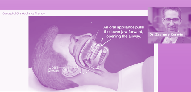
Mandibular advancement devices position the mandible forward during sleep to reduce upper-airway collapsibility. Qualified dentists have an established role in providing and managing oral appliance therapy following a medical diagnosis and prescription or referral.2,3
Research suggests that effective mandibular advancement therapy may reduce some jaw-closing muscle activity associated with respiratory arousals or oxygen-desaturation events in patients with OSA. However, investigators have also emphasized the need for further research, particularly in patients with confirmed comorbid sleep bruxism.8,10
A patient’s jaw-muscle activity may also be influenced by:
- Sleep arousals
- Autonomic activation
- Medications
- Alcohol or stimulants
- Other sleep disorders
- Individual motor patterns
For that reason, oral appliance therapy should be evaluated against its prescribed respiratory objective. Any change in RMMA, tooth grinding, or reported jaw activity should be considered a secondary observation unless it has been objectively assessed.
Why Technologist Workload May Matter
Not all attended sleep studies are conducted under identical conditions.
Ryan describes a growing operational challenge:
“I may be running three patients at once, and I’m the only technologist.”
A higher patient-to-technologist ratio does not automatically make a study inadequate. However, workload may affect how quickly a technologist can:
- Repair a failed lead
- Respond to signal loss
- Document unusual behavior
- Help capture body position
- Manage a patient’s immediate needs
- Review secondary channels
In a lower-ratio environment, the technologist may have more time to manage each study actively. In a higher-ratio setting, monitoring may become more reactive.
The final report should identify significant technical limitations. Dentists should review those notes instead of assuming that every PSG provides identical data quality.
What Dentists Should Review Before Oral Appliance Therapy
Dentists do not need to become sleep-study scorers to use the report effectively. However, a structured review can support case evaluation and communication with the treating physician.
Sleep-study review checklist
- Was the diagnosis based on PSG or home sleep apnea testing?
- Does the report provide AHI, REI, or both?
- How much sleep or monitoring time was recorded?
- Was adequate REM sleep captured?
- Did the patient spend meaningful time sleeping supine?
- Were events worse during REM or supine sleep?
- What was the lowest recorded oxygen saturation?
- Was cumulative time below a specified oxygen threshold reported?
- Were central or mixed events identified?
- Were significant cardiac findings noted?
- Did the report describe signal loss or technical limitations?
- Do the findings align with the patient’s symptoms and medical history?
- Has a qualified medical provider diagnosed OSA and recommended oral appliance therapy?
- What follow-up testing will be used to confirm treatment effectiveness?
Current standards emphasize coordinated care among the qualified dentist, diagnosing physician, and other members of the sleep-care team. Dentists evaluate the oral structures, provide and adjust the appliance, monitor dental effects, and refer the patient for objective assessment of treatment efficacy.2,3
When the Report and the Patient Do Not Match
A patient may report substantial symptoms despite a negative, mild, or technically limited home study. Conversely, a study may show clinically significant OSA in a patient who reports few symptoms.
Dentists should not resolve that discrepancy by making an independent medical diagnosis.
Instead, consider asking the physician:
- Was the home study technically adequate?
- Did the patient sleep for most of the monitoring period?
- Could insomnia have lowered the reported REI?
- Was positional or REM-related OSA a concern?
- Does the patient have a condition that makes PSG preferable?
- Would repeat or expanded testing be appropriate?
AASM guidance recommends PSG when a technically adequate home test is negative, inconclusive, or technically inadequate and clinical concern for OSA remains.1
Key Takeaway for Dentists
A sleep-study report is more than a severity category.
It reflects:
- The signals that were recorded
- Whether sleep was confirmed
- The stages and positions captured
- The quality of the recording
- The way respiratory events were calculated
- The interpreting physician’s clinical judgment
PSG provides detailed information about sleep stages, arousals, breathing, oxygen levels, body position, and related physiologic signals. Home sleep apnea testing provides greater convenience and access but generally offers less context.
Ryan’s advice is straightforward:
“You have to look at the whole picture.”
For dentists, that means reviewing more than AHI or REI. It also means considering sleep stage, position, oxygen findings, signal quality, and whether the results align with the patient’s clinical presentation.
When questions remain, collaboration with the diagnosing physician or sleep specialist is essential.
dentist’s role in dental sleep medicine
References
- Kapur VK, Auckley DH, Chowdhuri S, et al. Clinical practice guideline for diagnostic testing for adult obstructive sleep apnea: an American Academy of Sleep Medicine clinical practice guideline. J Clin Sleep Med.2017;13(3):479–504. doi:10.5664/jcsm.6506.
- Ramar K, Dort LC, Katz SG, et al. Clinical practice guideline for the treatment of obstructive sleep apnea and snoring with oral appliance therapy: an update for 2015. J Clin Sleep Med. 2015;11(7):773–827. doi:10.5664/jcsm.4858.
- Glick A, Chiang HK, Huynh N, Levine M. Dentistry’s role in the evidence-based management of adult obstructive sleep apnea: a narrative review. J Am Dent Assoc. 2026. doi:10.1016/j.adaj.2025.12.020.
- Lavigne GJ, Rompré PH, Poirier G, Huard H, Kato T, Montplaisir JY. Rhythmic masticatory muscle activity during sleep in humans. J Dent Res. 2001;80(2):443–448. doi:10.1177/00220345010800020801.
- Kato T, Rompré P, Montplaisir JY, Sessle BJ, Lavigne GJ. Sleep bruxism: an oromotor activity secondary to micro-arousal. J Dent Res. 2001;80(10):1940–1944. doi:10.1177/00220345010800101501.
- Kato T, Montplaisir JY, Guitard F, Sessle BJ, Lund JP, Lavigne GJ. Evidence that experimentally induced sleep bruxism is a consequence of transient arousal. J Dent Res. 2003;82(4):284–288. doi:10.1177/154405910308200408.
- Huynh N, Kato T, Rompré PH, et al. Sleep bruxism is associated with micro-arousals and an increase in cardiac sympathetic activity. J Sleep Res. 2006;15(3):339–346. doi:10.1111/j.1365-2869.2006.00536.x.
- Aarab G, Lobbezoo F, Hamburger HL, Naeije M. The effects of mandibular advancement appliance therapy on jaw-closing muscle activity in obstructive sleep apnea. J Oral Rehabil. 2020.
- Li D, Kuang B, Lobbezoo F, et al. Effects of continuous positive airway pressure and mandibular advancement appliance therapy on rhythmic masticatory muscle activity in adults with obstructive sleep apnea. 2023.
- Kuang B, Li D, Lobbezoo F, et al. The effects of mandibular advancement appliance therapy on jaw-closing muscle activities associated with oxygen desaturation and sleep arousal in obstructive sleep apnea. 2023.